Hemorrhage can result from trauma or vascular events in the brain tissue. Localizing and characterizing these types of lesions is key in forming a treatment plan and prognosis in dogs and cats. The physics of MRI and CT allow some staging of hemorrhage, and follow-up imaging can also be used to monitor resolution. Here is an excerpt from the Atlas of Small Animal CT and MRI from Section 6.5—Trauma, Hemorrhage, and Vascular Disorders.
Vascular disorders
Primary intracranial vascular disease is uncommon in cats and dogs, as compared to stroke disorders in people. Stroke occurs when blood flow to the brain is disrupted, causing ischemia and eventual brain cell death. Stroke is caused by either spontaneous vascular disruption, leading to hematoma formation, or from vascular occlusion, resulting in hemorrhagic or nonhemorrhagic infarction. Vascular occlusion (ischemic infarction) may be due to either in situ thrombus formation or obstructing emboli originating elsewhere. Hemorrhagic ischemic infarction occurs when the mural integrity of an occluded vessel is disrupted, secondarily leading to extravasation. It may be impossible to distinguish between hemorrhagic ischemic infarction and hematoma resulting from vascular disruption, as both will have similar imaging features. Most infarctions are arterial in origin, and although stroke from venous thrombosis is described in people, there are few comparable reports in veterinary medicine. The rostral and middle cerebral and the striate and rostral cerebellar arteries are the most commonly involved, and infarcts involving the cerebrum, thalamus/midbrain, and cerebellum have been reported. Infarcts are described as territorial when they involve a major intracranial vessel and lacunar when smaller penetrating vessels are obstructed. Underlying causes for stroke include atherosclerosis, hypertension, and diabetes in people, although these have not been confirmed as predisposing factors in veterinary patients.
Hematoma from vascular disruption
Hematomas from vascular disruption may occur as the result of vascular trauma or from spontaneous hemorrhage, as may occur with rupture of an intracranial vascular malformation. Imaging characteristics will vary depending on the size, location, and chronicity of the hematoma. Hematomas will generally appear as a hyperattenuating mass on unenhanced CT images, and there may be evidence of contrast enhancement if active bleeding (acute) or neovascularization (chronic) is present. MR imaging features will generally follow the scheme outlined in Table 2.4.1, although age can be ambiguous when multiple bleeding episodes occur over time. Secondary features of mass effect may include surrounding edema, midline shift, ventricular displacement and compression, and sulcus and gyrus effacement on both modalities.
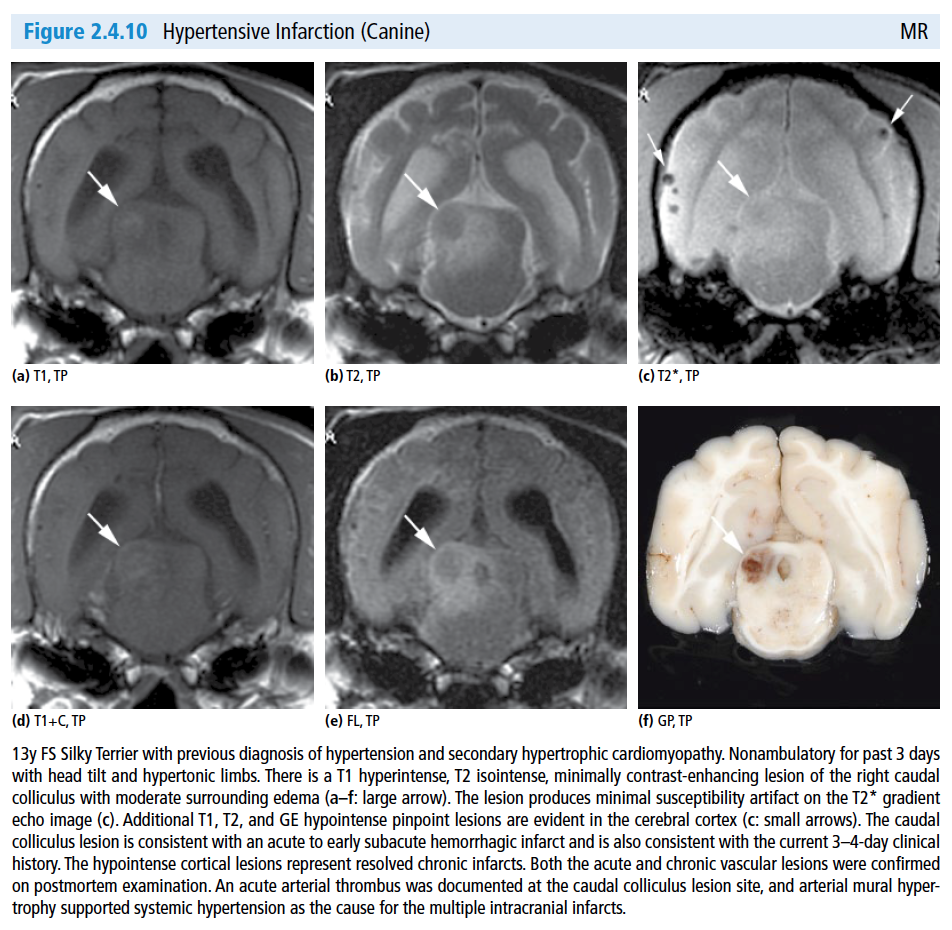
Hemorrhagic infarction
Hemorrhagic infarctions may not be distinguishable from hematoma caused by vascular disruption. Imaging features of hematoma described above are also applicable to hemorrhagic infarction.
Nonhemorrhagic Infarction
CT imaging features of nonhemorrhagic infarction may be subtle and include focal or regional hypoattenuation from edema and variable, but often minimal, mass effect.Nonhemorrhagic infarction may appear mildly T1 hypointense and T2 hyperintense with variable mass effect involving both gray and white matter on unenhanced MR images. Due to restricted water diffusion, ischemic regions of the brain will appear hyperintense on diffusion‐weighted images and hypointense on corresponding apparent diffusion coefficient (ADC) maps. Perfusion images may define specific regions of perfusion deficit, and magnetic resonance angiographic (MRA) images can reveal relative or absolute flow deficits in affected vessels. Gradient echo T2* images will display relatively little or no susceptibility effect.
Homework
Compare some of the images in your collection with the examples in this chapter to hone your interpretation skills. The Atlas of Small Animal CT and MRI is available through Amazon and has over 700 figures showing case examples to further your learning. Check it out if this article was helpful!
See you next time,
Allison Zwingenberger
Co-Author, Atlas of Small Animal CT and MRI

![]()
P.S.
Did you find this page via Twitter or a link from a friend?
Click here to subscribe today (at no charge) and get this excerpt course via email.
Of course, if you find it’s not for you, you can unsubscribe at any time with just a couple of clicks. We’ll never rent or share your information with anyone.